This week, we’re going over a new phage therapy case series report (Onsea et al. 2019). Although phage therapy to treat musculoskeletal infections (aka osteomyelitis) has historically been relatively common in Eastern Europe, most of the past studies have each been done differently, and have lacked relevant details regarding how they were done. This study aimed to take steps toward standardizing the process of phage therapy for osteomyelitis by detailing how a set of four patients were identified, treated and followed up with, along with details on the phages used.
(Here’s a previous Capsid & Tail post on how another group used phage therapy to treat this type of infection.)
The paper
The study was published last month in Viruses by Jolien Onsea of KU Leuven and the University Hospitals Leuven, alongside colleagues and collaborators at these institutes and at the Queen Astrid Military Hospital.
What are musculoskeletal infections?
Musculoskeletal infections are also referred to as osteomyelitis. Often, after implantation of orthopedic devices (e.g. prostheses), patients get infections. Often, bacteria are resistant to antibiotics, and biofilms are generally a huge problem.
What were the patients dealing with?
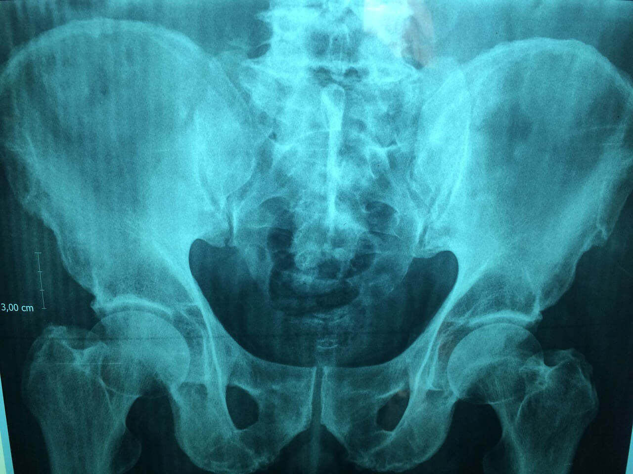
Four patients were found eligible for phage therapy. They were all facing poor prognoses (ie. likely amputation) because surgery and standard medical regimens had not helped. Three patients had chronic osteomyelitis of the femur. One patient had chronic osteomyelitis of the pelvis. They’d each suffered multiple infection relapses. One patient had received their diagnosis as early as 1984, another in 1995, another in 2015, and the other in 2017.
Which pathogens were cultured?
Multiple pathogens were isolated from the sites of infection. In two patients, Pseudomonas aeruginosa and Staphylococcus epidermidis were both isolated. One patient had Streptococcus agalactiae and Staphylococcus aureus, and another had Enterococcus faecalis.
Eligibility
A “multidisciplinary phage task force” (consisting of musculoskeletal surgeons, microbiologists, infectious disease specialists, plastic surgeons, and phage scientists) was assembled, and this team made the call about whether each patient was eligible for phage therapy.
To be eligible, patients had to fit the criteria declared in the Declaration of Helsinki (see our Capsid & Tail post here). This means that these patients had to have infections that weren’t being controlled by standard treatment options, and thus could qualify for unproven medical interventions (phage therapy).
Indeed, in all four cases, antibiotic therapy and surgery to remove the infected tissue had been tried, and had failed to clear the infections.
What needed to be done from an ethics standpoint?
Patients each gave their informed consent, and the hospital’s Ethical Committee gave consent for each patient to receive phage therapy.
Where did the phages come from?
Three patients received a phage cocktail called BFC1 (one S. aureus phage and two P. aeruginosa phages, all strictly lytic and well characterized) which was produced by the Queen Astrid Military Hospital. The phages in this cocktail were given a “genetic passport” by Sciensano, Belgium’s federal public health institute, as they were found not to contain toxins or antibiotic resistance genes.
The fourth patient received the Pyo phage cocktail from the Eliava Institute in Tbilisi, Georgia (contains multiple phages against Streptococcus, Staphylococcus, Proteus, Escherichia coli, P. aeruginosa, and Enterococcus, though the exact composition is not known).
Were patient strains first tested for phage susceptibility?
Yes. Three patients had Staphylococcus that was susceptible to BFC1. At least one of these patients also had P. aeruginosa that was resistant to this cocktail, but the team proceeded with the cocktail anyway based on its anti-Staphylococcus activity.
The fourth patient didn’t receive BFC1, as the cocktail doesn’t cover Enterococcus (the pathogen cultured from that patient). However, this patient’s E. faecalis was sensitive to the Pyo phage cocktail, so this patient received this cocktail instead (it was ordered from the Eliava).
How were phages applied?
Phages were given intraoperatively, by placing a draining system in close contact with the infected bone and rinsing with phage-containing solution. A sodium bicarbonate solution was applied just prior to the phage, to decrease the local acidity.
What was the dosage?
The BFC1 cocktail (used on three patients) was used at 10^7 PFU/mL in saline. Phages were given three times per day, for 7 to 10 days. Between 10-40 mL of phage solution was used to rinse the infected bone, and the phage was allowed to contact the site for 10 min (the drain was closed during this time). Before closing the wound, a gentamicin-impregnated collagen sponge soaked in phage solution was placed on the bone.
Were antibiotics used concurrently?
Yes. During phage therapy, each patient also received antibiotics (each received a different regimen, depending on their needs). The patients received antibiotics for six weeks to three months in total, even though the phages were only applied for 7-10 of these days.
How were the patients evaluated during and after phage therapy?
Each day, the patients’ clinical status was evaluated (this meant inspecting the wound, doing blood tests, checking general health status, and radiology). Blood was taken before surgery and on days 2, 4, 7, 10, 14, 28 post-surgery. Patient health was tracked for 8-16 months following phage therapy.
What was the outcome for the patients?
For three patients, no signs of infection recurrence were observed, and these patients are currently infection-free! (You can check out Figure 2 of the paper for before and after images, which are quite impressive).
One patient had to have more surgeries following phage therapy, to manage a bone defect. Eight months after phage therapy, another S. epidermidis strain was isolated from this patient. This strain had different antibiotic susceptibility compared to the first S. epidermidis strain isolated from this patient, so the team sequenced both. They confirmed the strains were not clonal (>8000 SNPs), meaning a new strain had come in to replace the first. Eventually, after additional surgeries and more antibiotics, this patient was also deemed infection-free.
Did the phages provoke inflammation?
Yes. C-reactive protein (CRP) and white blood cell (WBC) levels, markers of inflammation, were tracked in each patient over time. CRP levels increased in all patients during phage therapy, and WBC levels increased in two patients, but all levels returned to normal within a month following phage therapy in all patients.
Did the phages provoke anti-phage antibody production?
No. Serum samples were tested for their capacity to neutralize the administered phages, but anti-phage antibody production was not detected in any of the patients.
Were there any side effects?
No severe side effects were observed. The patient who got the Pyo cocktail had redness and pain during rinsing after 7 days of phage treatment, which may have resulted from a response to endotoxin present in the cocktail (Pyo cocktail is not free of endotoxin).
Summary
This paper describes the safe and successful use of phage therapy as an adjunct to antibiotics to treat severe polymicrobial musculoskeletal infections in four patients. Phages were given as pre-made, multi-target, intraoperatively-applied cocktails that were verified to lyse at least one of each patient’s isolated pathogens in advance.
Going forward
The “multidisciplinary phage task force” (infectious disease specialists, pharmacists, microbiologists, surgeons and phage scientists) assembled for this effort will continue to assess patients with musculoskeletal infections for phage therapy eligibility. Treatment plans will be developed for each, and data will be collected in a registry. The hope is that analyzing this data regularly will give insight into how to use phages to treat these types of patients safely and effectively.
Further reading
Other Capsid & Tail issues we’ve written on phage therapy case reports:


