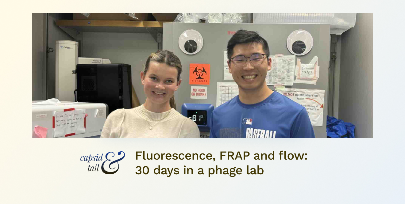
This past summer I had the incredible opportunity to spend the month working in the Bollyky Lab at Stanford University. As an incoming Senior in high school, my time was filled with new experiences and a deep dive into the world of microbiology and immunology. I worked with advanced lab techniques, including bacterial inoculation, plaque assays, fluorescently dyeing phages, flow cytometry, and fluorescence recovery after photobleaching (FRAP).
Basic Wet Lab Techniques
While I enjoyed being a part of experiments, I had a lot to learn regarding basic wet lab techniques. I had no prior experience working in a lab, so I had never heard of things such as top agar or plaque assays. I had never even used a pipette!
The very first thing my mentor (Tony Chang, a masters student in biology) taught me was the importance of sterilization and time efficiency. Sterilization is crucial because if something becomes unsterile, it can ruin the entire experiment. We have had multiple plaque assay experiments that went to waste all because of contamination. After seeing that, I became extra cautious when spraying my gloves and materials with alcohol, not wanting the time we spent on experiments to go to waste due to contamination. I also learned the vitality of keeping one’s hood organized in order to complete the experiment as quickly and accurately as possible. Oftentimes I would have two pipettes ready with different measurements so I wouldn’t have to stop my procedures. If I went too slow, the top agar would start to dry resulting in clumpy plates. Time management and sterilization are vital aspects of every experiment and their success.
Plaque Assays
Plaque assays were a central part of our research and the part at which I spent most of my time. They are used to measure the ability of phages to infect bacterial cells. I first added bacteria to the top agar solution. I then completed the sterile dilutions by slowly diluting each phage with phosphate-buffered saline (PBS). I put a small amount of each dilution on the agar plates and left them overnight. As the phages infected and killed the bacteria, they formed clear spots called plaques. By counting these plaques, we could quantify the phage concentration and their infectivity.
The plaque assays required proper sterilization and time management to ensure accurate results. Oftentimes we would have to repeat the assays multiple times before getting results due to contamination or inconsistencies. Overall, it was fascinating to see the direct impact of phages on bacterial populations, providing valuable data for our research.
In addition to plaque assays, I spent a lot of time inoculating bacteria and making bacteria cultures. To create bacteria cultures, I took an inoculation loop and spread bacteria on agar plates and then incubated the plates. Inoculating bacteria involved introducing bacteria taken from bacteria cultures I previously made and introducing it into a growth medium under sterile conditions to cultivate them for experiments. We then used this bacteria for other experiments including top agar for plaque assays. I learned to handle pipettes and inoculating loops with precision, transferring tiny volumes of bacteria to agar plates or liquid media. It was a delicate balance between maintaining sterility and working efficiently, and I quickly developed a steady hand and an appreciation for the meticulous nature of microbiological work.
Fluorescently Dyeing Phages
My initial project in the lab was the fluorescent dyeing of phages. Our first objective was to determine if the lytic phage PAML would express fluorescent dye, allowing it to be visualized through a microscope. We then wanted to observe how the phage would diffuse in a biofilm setting. The best way to observe this was through Fluorescence Recovery after Photobleaching or FRAP. We used a conjugate of the dye Alexa Fluor 488 and a lytic phage called PAML-31-1 (PAML for short).
The experimental setup included four different wells in a 384 well plate for ease of multiple experiments:
- well 1 contained dye only,
- well 2 combined PAML and dye,
- well 3 included PAML, dye, and a polymer to mimic biofilm conditions,
- well 4 comprised PAML, dye, polymer, and Pf, a lysogenic phage (because the Bollyky lab previous discovered how this lysogenic phage can integrate itself into the bacterial host Pseudomonas aeruginosa genome and provide a structural element that contributes to the bacteria’s overall virulence).
Flow cytometry analysis was utilized to reveal that the dye was successfully taken up by the phage. To finalize our results, we used fluorescence recovery after photobleaching (FRAP) to test the diffusion rate of the dye within the phages. This technique involves bleaching the dye with a laser and measuring the amount of time it takes for the fluorescence to return. This provides insights into the dye’s mobility within the phages. This experiment was a fascinating introduction to my time in the lab, and became the central focus of my research experience.
Reflection
My month at one of Stanford’s infectious disease labs was filled with learning, discovery, and hands-on experiments. However, the part that made this experience so phenomenal was Dr. Paul Bollyky and my mentor, Tony Chang. Without their patience and constant support, I would have never had the opportunity to learn and participate in the lab as much as I did.
As I look forward to a future in medicine, I will look back at this experience and remember all of the invaluable lessons and knowledge I gained this summer working in the Bollyky Lab. This month leaves me with a deeper understanding of the importance of research and a strong desire to be part of a community that is constantly asking questions and making new impactful discoveries in science and ultimately healthcare.
